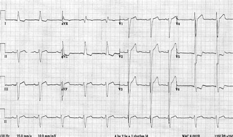
lv hypertrophy ecg | signs of lvh on ecg lv hypertrophy ecg The left ventricle hypertrophies in response to pressure overload secondary to conditions such as aortic stenosis and hypertension. This results in increased R wave amplitude in the left-sided ECG leads (I, aVL and V4-6) and increased S wave depth in the right-sided . Edit - As for XP boosts, i save them for new champs. Get a champ to lv13 and do any 3* or higher adventure with the cosmic blessing to get them to lv20+ straightaway. If you have surplus cosmic blessing you can use them on lv 25 champs and do Asol adventure, this should get them to lv 30.
0 · what is lvh on ecg
1 · signs of lvh on ecg
2 · lvh with repolarization abnormality ecg
3 · lv hypertrophy ecg criteria
4 · left ventricular hypertrophy with repolarization abnormality
5 · left ventricular hypertrophy life in the fast lane
6 · ecg showing lvh
7 · ecg in left ventricular hypertrophy
We have 1 Eyedro EHEM manual available for free PDF download: Product Manual. Eyedro EHEM Product Manual (45 pages) ELECTRICITY MONITORING PRODUCTS. Brand: Eyedro | Category: Measuring Instruments | Size: 3.36 MB. Table of Contents. 2. Introduction. 4. Overview. 4. Important Safety Information. 5. Box Contents (by Product) .
The left ventricle hypertrophies in response to pressure overload secondary to conditions such as aortic stenosis and hypertension. This results in increased R wave amplitude in the left-sided ECG leads (I, aVL and V4-6) and increased S wave depth in the right-sided .

The ECG Made Practical 7e, 2019; Kühn P, Lang C, Wiesbauer F. ECG Mastery: .
ECG Pearl. There are no universally accepted criteria for diagnosing RVH in .
Also known as: Left Atrial Enlargement (LAE), Left atrial hypertrophy (LAH), left .
The delay between activation of the RV and LV produces the characteristic “M .
The ECG Made Practical 7e, 2019; Kühn P, Lang C, Wiesbauer F. ECG Mastery: .Kühn P, Lang C, Wiesbauer F. ECG Mastery: The Simplest Way to Learn the .Learn how to interpret ECG changes in LVH, such as large R-waves in left-sided leads and deep S-waves in right-sided leads. Find out the common causes, . The left ventricle hypertrophies in response to pressure overload secondary to conditions such as aortic stenosis and hypertension. This results in increased R wave .
The most common causes of left ventricular hypertrophy are aortic stenosis, aortic regurgitation, hypertension, cardiomyopathy and coarctation of the aorta. There are several ECG indexes, . Left ventricular hypertrophy changes the structure of the heart and how the heart works. The thickened left ventricle becomes weak and stiff. This prevents the lower left heart . Left ventricular hypertrophy (LVH) refers to an increase in the size of myocardial fibers in the main cardiac pumping chamber. Such hypertrophy is usually the response to a .
Electrocardiogram. Also called an ECG or EKG, this quick and painless test measures the electrical activity of the heart. During an ECG, sensors called electrodes are . CONTENTS LAD (left axis deviation) LAHB (left anterior hemiblock) iLBBB (incomplete left bundle branch block) LVH (left ventricular hypertrophy) Diagnostic criteria . According to the American Society of Echocardiography and/European Association of Cardiovascular Imaging, LVH is defined as an increased left ventricular mass index (LVMI) .Left ventricular hypertrophy can be diagnosed on ECG with good specificity. When the myocardium is hypertrophied, there is a larger mass of myocardium for electrical activation to .
Interval between QRS and R-wave peak in V5 or V6 ≥ 0.05 second. 1. Sokolow-Lyon. V1 S wave + V5 or V6 R wave ≥ 35 mm. or. aVL R wave ≥ 11 mm. N/A. ECG = electrocardiography; LVH .
louis vuitton swimsuit china
Left ventricular hypertrophy with increased precordial voltages and non-specific ST segment and T-wave abnormalities. Deep, narrow (“dagger-like”) Q waves in lateral (I, aVL, .
The left ventricle hypertrophies in response to pressure overload secondary to conditions such as aortic stenosis and hypertension. This results in increased R wave .The most common causes of left ventricular hypertrophy are aortic stenosis, aortic regurgitation, hypertension, cardiomyopathy and coarctation of the aorta. There are several ECG indexes, .
what is lvh on ecg
Left ventricular hypertrophy changes the structure of the heart and how the heart works. The thickened left ventricle becomes weak and stiff. This prevents the lower left heart . Left ventricular hypertrophy (LVH) refers to an increase in the size of myocardial fibers in the main cardiac pumping chamber. Such hypertrophy is usually the response to a . Electrocardiogram. Also called an ECG or EKG, this quick and painless test measures the electrical activity of the heart. During an ECG, sensors called electrodes are .
CONTENTS LAD (left axis deviation) LAHB (left anterior hemiblock) iLBBB (incomplete left bundle branch block) LVH (left ventricular hypertrophy) Diagnostic criteria . According to the American Society of Echocardiography and/European Association of Cardiovascular Imaging, LVH is defined as an increased left ventricular mass index (LVMI) .Left ventricular hypertrophy can be diagnosed on ECG with good specificity. When the myocardium is hypertrophied, there is a larger mass of myocardium for electrical activation to .Interval between QRS and R-wave peak in V5 or V6 ≥ 0.05 second. 1. Sokolow-Lyon. V1 S wave + V5 or V6 R wave ≥ 35 mm. or. aVL R wave ≥ 11 mm. N/A. ECG = electrocardiography; LVH .
signs of lvh on ecg
lvh with repolarization abnormality ecg
You can purchase FABER CASTELL BALLPOINT STICK 246629 LV7 BLUE here at nationalbookstore.com
lv hypertrophy ecg|signs of lvh on ecg


